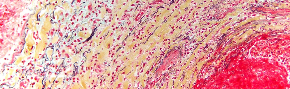
Show Commentary
PNEUMOCYSTIS PNEUMOCYSTIS (2013)
Expanded commentary:
Pneumocystis pneumonia is a common cause of death in patients whose immune systems are weakened by chemotherapy, bone marrow transplantation, or (most commonly) infection with human immunodeficiency virus (HIV). Pneumocystis is a yeast-like fungus that was rarely diagnosed before the onset of the acquired immune deficiency syndrome (AIDS) epidemic. A paper published in 1982 in the
Morbidity and Mortality Weekly Report published by the Centers for Disease Control noted an unexplained increase in the incidence in Kaposi’s sarcoma (a cancer associated with AIDS that causes reddish-purple skin lesions) and Pneumocystis pneumonia in residents of southern California. After several case studies of the disease, the paper rather poignantly notes: “All the above observations suggest the possibility of a cellular-immune dysfunction related to a common exposure that predisposes individuals to opportunistic infections such as pneumocystis and candidiasis”. Marked by shortness of breath, persistent fever, night sweats, and unintended weight loss, Pneumocystis
pneumonia is now less commonly diagnosed due to the availability of antiretroviral medications to treat HIV infection. This photomicrograph illustrates Pneumocystis infection in an immunocompromised patient.
Pneumocystis pneumonia is characterized histologically by the presence of tiny, clustered, cup-shaped organisms in the spaces within the lung. The organisms are highlighted in black against a green background by the application of the Gömöri methenamine silver (GMS) stain. GMS is a common histological stain used to detect the presence of organisms, particularly fungi, by blackening components of the cell wall while coloring elements of normal human tissue green. The presence of Pneumocystis
pneumonia in a post-mortem specimen strongly suggests that a weakened immune system caused or contributed to the patient’s death. An explanation for the immunocompromised state must be found before an autopsy report or death certificate can be properly completed.
TRICHROME STAIN OF SMALL BOWEL #1 (2013) and #2 (2013)
Short explanation:
The Masson’s trichrome stain highlights tissue subtypes in small bowel.
Expanded commentary:
When a pathologist requests a special stain, like Masson’s trichrome, on a piece of tissue, the histologist will often include an extra slide when the case is returned to the physician for interpretation. The extra slide is a control – a piece of tissue that is used again and again to test that the stain actually worked! Both of these images were captured from a control slide of small bowel used to make sure the trichrome stain was in good order.
TATTOO (2013)/TATTOO 400X (2013)/TATTOO RED PIGMENT(2014)
Short explanation:
Tattoo pigment is visible under the microscopic in the dermis, or the deep layer of the skin, and retains its color at high magnification.
Expanded commentary:
An old saying states that “every generation thinks it invented sex”; apparently, every generation also thinks it invented tattoos. In point of fact, the act of tattooing – or intentionally depositing dye in the deeper layers of the skin to create a permanent design – has been part of worldwide artistic and cultural expression for more than 5000 years. Tattoos have been noted on Iron Age bodies pulled from the ice of the Alps, ancient Peruvian mummies, and (more recently) British royalty. The social acceptability of tattoos has varied widely throughout history; although outlawed in the Old Testament, tattoos indicating religious devotion were popular among early Christians. Tattoos now openly memorialize loved ones, indicate group membership, mark time behind bars, or serve as landmarks for radiation treatments.
Under the microscope, tattoo pigment is found in the dermis, the deeper layer of the skin.
Macrophages and histiocytes (two types of cells found in the skin) are unable to completely process and dispose of pigment granules. At high magnification, individual fragments of tattoo dye are visible, as are their color. Though initially distinct, the body’s attempt to rid itself of the pigment is ongoing and progressive, and some tattoos fade and blur with time. In particular, repeated bouts of sun exposure will lessen the sharpness and contrast of a tattoo.
A small fraction of the public regrets their choice of tattoo and laser removal is sometimes a solution. Removal works by a process called selective thermolysis – that is, pigment particles are heated, causing them to break into smaller pieces, which allows cells to engulf and clear them. Dyes comprised of smaller particles at the time of application are more difficult to fragment with heat and remove.
Death investigators largely use tattoos to determine an unknown decedent’s identity through comparison with pre-mortem photographs or descriptions from loved ones. Counterintuitively, tattoos are often more visible in slightly putrefied bodies. One of the first changes of decomposition is the loss of the outer layer of the skin, making old tattoos appear bright and sharp once again.
AMYLOIDOSIS – CONGO RED (2013) and BIFREFRINGENCE (2014)
Short explanation:
Pathologists use a special stain called Congo Red and polarized light to diagnose amyloidosis, or a disease of abnormal protein deposition.
Expanded commentary:
Amyloidosis is a family of diseases in which abnormal proteins, called amyloids, are deposited in human tissue and disrupt normal organ function. The cause of amyloidosis is, as yet, poorly understood. Symptoms of amyloidosis correspond to the organs most markedly affected by the disease, but can include abnormal heart function, splenic rupture, enlargement of the tongue, vomiting, and/or hemorrhage from the respiratory or gastrointestinal tract. Diagnosis results from the microscopic examination of tissue treated with a special stain known as Congo Red, which colors amyloids a distinctive pink or red hue. As the manifestations of the disease can be varied and non-specific, the diagnosis of amyloidosis is not infrequently made after post-mortem examination. These panels highlight amyloid deposits around blood vessels using under normal and polarized light sources.
Invented in 1883 in Germany as a coloring agent for textiles, the Congo Red special stain has a special affinity for the abnormal proteins present in amyloidosis. The dye was dubbed “Congo Red” not to reflect its origin (it was entirely manufactured from waste products of Germany’s coal gas and steel industries) but instead as a marketing ploy, intended to highlight its rarity and exoticism. Congo Red turned out to be an impractical dye for clothing but testing on histologic specimens revealed it to be an avid binding agent of amyloid proteins. Later experiments with polarized light revealed that tissue specimens with abnormal amyloid protein that were stained with Congo Red glowed apple-green under specialized conditions. Since the early 20th century, pathologists have relied on Congo Red to visualize the sometimes subtle signs of amyloidosis in human tissue.
CRACK LUNG (2013)
Short explanation:
White blood cells stuffed with products of combustion fill the lungs of a chronic user of crack cocaine.
Expanded commentary:
“Complications of chronic substance abuse” is a phrase that appears with relative frequency on death certificates. While the short-term risks of alcohol abuse or the use of illicit drugs are well-documented, the effects of chronic substance abuse on an individual’s health receive far less press. A negative post-mortem toxicology report does not negate the pathological effects of years of substance abuse. The ravages of long-term drug abuse are easily seen at high magnification. Some presentations are so striking as to have earned themselves a moniker – in this case, “crack lung”. Crack lung is a syndrome of lung damage marked by the presence of macrophages (white blood cells adapted to engulf and destroy bacteria, dead or dying cells, cellular debris, and foreign material) filled with brownish-black foreign material within the airspaces of the lung. The brownish-black pigment generated from crack use remains within the macrophages of the lung indefinitely and is visible microscopically. The presence of pigment-laden macrophages results from inhalation of products of combustion during crack use. Unlike cigarettes, crack pipes lack a filter and users often breathe in large fragments of partially burned material. Macrophages patrol the bloodstream and tissues and engulf such material. This photomicrograph is representative of the lungs of a long-term abuser of crack cocaine.
Physiologically, the inhalation of heated crack cocaine (cocaine treated with sodium bicarbonate, which lowers the temperature of vaporization and makes it smokable) leads to marked increases in heart rate, blood pressure, and vascular constriction. Using a hot crack pipe may cause burns of the fingers (sometimes severe enough to obliterate the fingerprints), lips, oral cavity, and airway. Used chronically, the physiological effects of crack – so named for the crackling noises producing during its manufacture – are progressive and potentially deadly. Microscopic findings consistent with crack lung strongly suggest a history of chronic substance abuse to the astute pathologist and should spur histologic examination of the heart for thickening of the intramyocardial vessels, myocyte hypertrophy, and accelerated atherosclerosis that may have caused disability and death long after the cessation of drug use.
CRYPTOCOCCUS (2014)
Short explanation:
Cryptococcus is a commonly found pathogen that infects patients with weakened immune systems.
Expanded commentary:
Cryptococcus neoformans is a yeast-like fungus that gained prominence in the medical community due to its tendency to sicken the immunocompromised. Present in soil world-wide, the opportunistic pathogen (an organism that preys on patients with weakened immune system) enters a host through inhalation of air-borne spores and has the potential to cause cryptococcal meningitis by infecting the membranes surrounding the brain and spinal cord or cryptococcosis, an infection of multiple tissues. Cryptococcus is not transmissible from person-to-person and its long incubation period before causing clinically-significant symptoms often creates difficulty in pinpointing the source of the fungus. The name “Cryptococcus” means “hidden sphere” and pathologists employ special histological stains to visualize and identify the organism within tissue. Each round fungal organism is surrounded by a colorless mixture of protein and carbohydrates. The application of a mucin stain colors the capsule of Cryptococcus red and facilitates its microscopic identification. This photomicrograph shows cryptococcal infection of the liver, consistent with cryptococcosis.
ETHYLENE GLYCOL (2010)
Short explanation:
Ethylene glycol, also known as antifreeze, can be fatal if ingested. Breakdown products of antifreeze can be identified as crystals in the victim’s kidney at high magnification.
Expanded commentary:
Ethylene glycol, or antifreeze, is an organic compound commonly used to lower the freezing point of an antifreeze/water mixture. Odorless, colorless, and sweet-tasting, ingested antifreeze is potentially toxic in small quantities. The culprit in antifreeze poisoning is not ethylene glycol itself but instead its metabolites (or breakdown products), glycolic acid and oxalic acid. Ingestion of antifreeze, whether homicidal, suicidal, or accidental, offers a slow and painful death if medical intervention is not sought. The initial symptoms of antifreeze poisoning include central nervous system depression manifested through slurred speech, incoherence, lack of coordination, and vomiting. After 12 hours, pH abnormalities and cardiovascular dysfunction result. Approximately 36 hours after ingestion normal kidney function ceases
FAT EMBOLI – OIL RED O (2010)
Short explanation:
Fractures can cause displacement of bone marrow into the bloodstream and eventually, the lungs, called fat emboli.
Expanded commentary:
Fat emboli are a common consequence of blunt impact injuries, bone fractures, and cardiopulmonary resuscitation (CPR). Fat is a component of bone marrow, the tissue within the pelvis, sternum, vertebrae, and ribs of adults that produces the cellular elements of blood. Bony fractures may cause the embolization (inappropriate movement) of bone marrow into the bloodstream. The vast majority of fat emboli are asymptomatic and without consequence; however, a small percentage are clinically relevant and potentially fatal. Known as fat embolism syndrome, this phenomenon occurs 24 to 72 hours after blunt impact injury and is characterized by sudden-onset shortness of breath, a fine red rash, mental status changes and, in some cases, death. The precise mechanism with which fat embolism syndrome causes death is, as yet, unclear.
Oil Red O is a fat-soluble dye that colors fat (normally white on histologic section) a brilliant red hue. The application of Oil Red O to lung tissue identifies and highlights the fat cells in intravascular bone marrow emboli if they have migrated to the lungs. The presence of fat emboli in the lungs in conjunction with a clinical history of skeletal fractures and sudden death is consistent with fat emboli syndrome.
GUNSHOT WOUND (2010)
Short explanation:
Microscopic examination of the skin around gunshot entrance wounds may reveal the presence of soot, hemorrhage, and bone fragments.
Expanded commentary:
The microscopic identification of soot surrounding and within a gunshot wound is used by forensic pathologists to narrow the range of fire – that is, the distance between the barrel of the gun and the victim. While it’s well-known that firing a gun discharges a bullet, less obvious are the other substances which also exit the barrel. The effects of flame, soot, and unburned gunpowder allow a skilled pathologist to classify the entrance as a contact (in which the muzzle is pressed into the skin), near-contact, intermediate, or distant wound. Contact wounds often demonstrate thermal injuries of the wound edge and soot in the bullet track, whereas near-contact wounds will deposit soot (burned gunpowder) adjacent to the gunshot wound. As the distance between the end of the gun barrel and the
victim increases, the likelihood that fragments of unburned gunpowder (called “stippling”) will mark the skin surrounding the gunshot wound increases. Distant gunshot wounds will demonstrate no soot, stippling, or thermal injury to the edges of the wound. The blackening of skin associated with thermal injury and soot deposition can be difficult to distinguish with the naked eye, but are distinctively different under the microscope. This image shows the presence of brownish-black soot within the gunshot wound, consistent with a contact wound; also identifiable are skin, red blood cells consistent with acute hemorrhage, and a small bone fragment.
INTRAVENOUS DRUG ABUSE, LIGHT (2014) and DARK (2014)
Short explanation:
Intravenous drug abuse may lead to fillers and adulterants like talc lodging in the blood vessels of the lung; the deposits are especially visible under polarized light.
Expanded commentary:
Semi-synthetic opioids like oxycodone, fentanyl, hydrocodone, hydromorphone, and methadone are derivatives of thebaine, a component of the poppies from which opium is produced. Introduced in 1917 as painkillers, the abuse of opioids is now a public health issue of epidemic proportions.
Chronic intravenous use of prescription medications formulated for oral administration can be diagnosed through histologic examination of the lungs. Prescription medications in tablet form are comprised of an active ingredient and bulking agents such as cellulose, starch, or magnesium trisilicate (better known as talc). Introduction of crushed prescription medications dissolved in water into the vascular system may cause pulmonary deposition of fillers within the spaces around the blood vessels and the formation of granulomas (a collection of immune cells that attempts to wall off foreign material). Although such granulomas are commonly found in the lungs of prescription drug abusers, they are rarely symptomatic. Polarized lights highlights birefringent crystals that may identify the foreign material present: starch generates bright patterns in a Maltese cross formation, while talc deposits cause needle-shaped or plate-like crystals. Identification of foreign material around the blood vessels within the lung indicates chronic intravenous substance abuse.
LUNG CARCINOMA (2014)
Short explanation:
Lung cancer cells surround a fragment of cartilage.
Expanded commentary:
Individuals who die of cancer are often under the care of a physician who will sign their death certificate, and as such don’t require post-mortem examinations at the medical examiner’s office. However, pathologists occasionally encounter natural deaths due to cancer that were previously undiagnosed for whatever reason – late presentation, inability to get care, or masking of symptoms due to other issues. This image shows the relative disorganization of cancer cells due to unchecked growth. Advanced tumors may show central necrosis – that is, death of part of a tumor mass – because the lesion has grown faster than the body’s blood supply.
PENTACHROME #2 (2017)
Short explanation:
The pentachrome stain uses five colors to highlight elements of human tissue and is especially useful in the evaluation of cardiovascular specimens, like this section of artery wall.
Expanded commentary:
Movat’s Pentachrome stain generates some of the most diverse results in histology. As the name suggests, the stain is a combination of five dyes, each with an affinity for a different tissue element; depending on the source and tissue constituents found in the tissue on the slide, the images produced by the pentachrome stain can vary wildly, from a complex interplay of yellow, black and red in cardiac specimens to a near-metallic gold and rose in cardiac nerves or the brilliant red and blue of intestinal tissue. Pathologists rely on pentachrome to reveal subtle, early changes of disease that can’t be seen with the standard pink-purple combination of hematoxylin and eosin.
Developed in 1955 by Canadian Henry Zoltan Movat (1923–1995)1 to distinguish elements of connective tissue and modified in 1972 by H.K. Russell Jr.2 to shorten the staining time and improve reliability, pentachrome’s power lies in its ability to simultaneously highlight multiple subcellular elements, revealing changes of age, disease, and development. Today the pentachrome stain is commonly used by cardiac pathologists to see the relative proportions of normal and abnormal tissues in biopsy and autopsy samples. If you’ve ever pondered the deep philosophical question of what you’re made of, pentachrome has the answer: connective tissue, mostly.
1 Movat, HZ (1955). “Demonstration of all connective tissue elements in a single section; pentachrome stains”. AMA Archives of Pathology. 60 (3): 289–95.
2 Russell Jr, HK (1972). “A modification of Movat’s pentachrome stain”. Archives of Pathology. 94 (2): 187–91
SOOT IN AIRWAY (2017)
Short explanation:
Black soot visible on the surface of the airway indicates the decedent was alive at the time of the fire.
Expanded commentary:
The presence of soot on the surface of the airway answers a critical question in death investigation: did the decedent die because they burned, or did they burn because they died? In other words, was the individual already dead at the time the fire was set, or did they expire due to thermal injuries and/or smoke inhalation?
Products of combustion do not move passively into the deep channels of the lung during a fire. If the pathologist is able to identify soot on the surface of the bronchi (the main entrances to the lungs) or any of the branching channels through which air moves throughout the respiratory system, she or he can say definitively if the victim was alive during the conflagration.
THREE CARDIAC NERVES (2015)
Short explanation:
Cross-section of a nerve bundle of the soft tissue of the heart treated with a pentachrome stain.
Expanded commentary:
Movat’s Pentachrome stain generates some of the most diverse results in histology. As the name suggests, the stain is a combination of five dyes, each with an affinity for a different tissue element; depending on the source and tissue constituents found in the tissue on the slide, the images produced by the pentachrome stain can vary wildly, from a complex interplay of yellow, black and red in cardiac specimens to a near-metallic gold and rose in cardiac nerves or the brilliant red and blue of intestinal tissue. Pathologists rely on pentachrome to reveal subtle, early changes of disease that can’t be seen with the standard pink-purple combination of hematoxylin and eosin.
Developed in 1955 by Canadian Henry Zoltan Movat (1923–1995)3 to distinguish elements of connective tissue and modified in 1972 by H.K. Russell Jr.4 to shorten the staining time and improve reliability, pentachrome’s power lies in its ability to simultaneously highlight multiple subcellular elements, revealing changes of age, disease, and development. Today the pentachrome stain is commonly used by cardiac pathologists to see the relative proportions of normal and abnormal tissues in biopsy and autopsy samples. If you’ve ever pondered the deep philosophical question of what you’re made of, pentachrome has the answer: connective tissue, mostly.
3 Movat, HZ (1955). “Demonstration of all connective tissue elements in a single section; pentachrome stains”. AMA Archives of Pathology. 60 (3): 289–95.
4 Russell Jr, HK (1972). “A modification of Movat’s pentachrome stain”. Archives of Pathology. 94 (2): 187–91.
MICROSTEATOSIS (2014) AND BILE LAKES (2014)
Short explanation:
Microsteatosis (the presence of tiny fat globules) and bile retention are common and benign findings in the liver.
Expanded commentary:
Quirks of human microanatomy are frequently identified upon post-mortem histological examination. Among the most important lessons in pathology training is the recognition of benign entities that, while interesting, had little or no clinical significance in life. Correct identification and placement of such findings in their proper context is crucial to the determination of cause and manner of death.
One of the functions of the cells of the liver is to produce bile, which aids in digestion of fats. Formed from bilirubin, conjugated bile salts, cholesterol, and phospholipids, bile flows through a series of ducts that terminate in the small bowel. (Excess bile that is not immediately needed is stored in the gallbladder.) Obstruction of the flow of bile can lead to the formation of bile lakes, or areas of pooling within the liver, caused by metabolic or autoimmune disease, malignancy, or (most commonly) gallstones. Bile ranges in color from light amber to dark greenish-black, and such bile lakes are not infrequently encountered in post-mortem liver histology.
The liver is the primary organ for fat metabolism, and retention of fat globules is a common finding in post-mortem histology. Known as steatosis, accumulation of excess fat in the liver has many causes including obesity, alcoholism, infection with hepatitis C, and diabetes mellitus. Histological processing of liver tissue dissolves fat leaving clear spaces within the tissue. However, dyes that bind fat can be applied to make clear the extent and cellular location of the steatosis. In this example, the Oil Red O special stain has been applied to liver tissue to highlight microsteatosis, or the presence of fat particles within liver cells that do not distort the cell structure.
GOMORI TRICHROME STAIN OF ARTERY (2014) and
ELASTIC STAIN OF ARTERY (2014)
Short explanation:
Special stains highlight connective and elastic tissue elements in a blood vessel wall.
Expanded commentary:
The anatomy of blood vessels is highlighted by the use of the elastic and trichrome stains. Arteries, the vessels that deliver oxygenated blood from the heart to organs and tissues, contain the protein elastin within their walls, allowing them to stretch in response to the pressure wave generated by each heartbeat. The elastic stain colors elastin fibers in the artery wall black. Reductions or disruptions in the elastic layer of arteries leave patients at risk for internal hemorrhages due to aortic dissections or aneurysms, as in Marfan disease.
The Gömöri Trichrome stain is a mixture of dyes that, in combination, vividly differentiates tissue types from one another. The blue-green color of connective tissue contrasts with the reddish-purple of muscle tissue and allows the pathologist to evaluate the relative proportions of tissue types within the vessel wall.
GLOMERULUS (2013) and NERVE (2013)
Short explanation:
There are images of normal structures in the kidney (Glomerulus) and muscle (Nerve).
Expanded commentary:
Despite the frequency with which death investigation is depicted in the media, few (if any) on-screen forensic scientists appear to use microscopy as a tool to determine cause and manner of death. The reality is that microscopy is one of the pillars on which a good-quality investigation may rest and can reveal information that is otherwise unavailable. Pathologists use histology (the study of the microscopic structures of human tissues) to diagnose natural disease, identify injuries, or evaluate the presence and meaning of foreign material like soot in a gunshot wound or bulking agents used in the preparation of illicit drugs. The application of polarized light may offer an additional histological perspective.
Because thin slices of tissue are colorless under the microscope, dyes that bind and delineate cell structures are added during the generation and processing of glass slides. The standard stains used by almost all pathologists are hematoxylin and eosin, which color parts of the cell pink and purple. Normal microanatomic structures are commonly encountered during microscopic examination, like the functional unit of the kidney as shown in Glomerulus or the bundles of sensory fibers caught in cross- section in Nerve. Other dyes – known categorically as “special stains” – can be used to highlight aspects of cellular morphology, identify foreign material or deposits of disease-causing proteins, or distinguish cell types from one another.
THYROIDIZATION OF KIDNEY (2014)
Short explanation:
This image shows the changes of a kidney affected by end-stage renal disease, or kidney failure. Histologically, non-functioning kidneys closely resemble thyroid tissue – hence, the term “thyroidization of the kidney”.
PENTACHROME OF CARTILAGE (2019) and CHRISTMAS COLON (2019)
Short explanation:
Sections of cartilage and colon treated with the pentachrome stain.
VESSEL WALL SERIES
PAS STAIN (2015)
ELASTIC STAIN (2019)
TRICHROME STAIN (2015)
VVG STAIN (2019)
Short explanation:
This is a section of blood vessel wall treated with four different special stains to highlight different elements of the tissue
CORONARY ARTERY SERIES
(Dissection #1, Dissection #2, Recanalization, Pinpoint stenosis (2023), Pentachrome)
Expanded commentary:
The coronary arteries are three small vessels that supply blood and oxygen to the heart muscle itself. As they are critical to life, forensic pathologists take a special interest in the three main coronary arteries (the left anterior descending, the right, and the left circumflex) and their branches. Coronary artery stenosis is better known as “heart disease” and refers to the buildup of fats and cholesterols on the inner surface of the vessels, partially or completely blocking blood flow to the heart. The vessels also sometimes undergo calcification with age, better known as “hardening of the arteries”. Full or partial cross-sections of the arteries show the narrowing and calcification of vessels. Coronary artery dissections occur when a small tear of the inner surface of the vessels allows blood to accumulate in the space between the layers of the artery wall, sometimes compressing the main channel and preventing blood from reaching the heart muscle itself. Dissections are caused by stress on the wall of the coronary artery and can be associated with drug use, pregnancy, and trauma.
Recanalization of a coronary artery occurs when small channels form to bridge a blocked area.